One Unreported Anesthesia Protocol Slowed a Whole-Brain Calcium Imaging Atlas
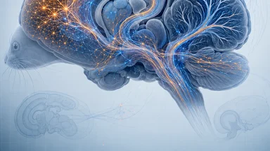
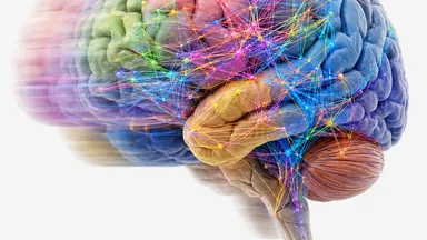
Whole-brain calcium imaging promised a functional connectome—a map of every neuron's activity across the brain. But one lab's dataset showed a troubling pattern: deep structures like the hippocampus were nearly silent, while cortical regions lit up normally. The project stalled for nearly a year. The culprit was not a hardware failure or a software bug, but a single variable that no one had calibrated: the anesthesia protocol.
Why an Atlas of 100,000 Neurons Ground to a Halt
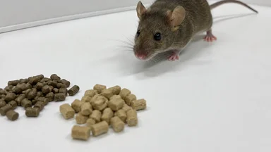
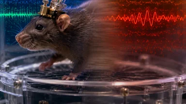
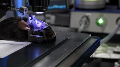
The goal was ambitious: image calcium transients from roughly 100,000 neurons across multiple brain regions in head-fixed mice, while the animals performed a simple behavioral task. The team at a mid-sized neuroscience institute had invested heavily in a custom two-photon mesoscope, capable of recording from thousands of neurons simultaneously. Early runs looked promising. Cortical regions showed robust activity patterns. But when the team examined subcortical areas—the hippocampus, thalamus, and basal forebrain—they saw something puzzling. Calcium transients were barely detectable. The hippocampus, a region known for place-cell activity and sharp-wave ripples, appeared almost inert.
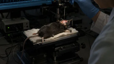
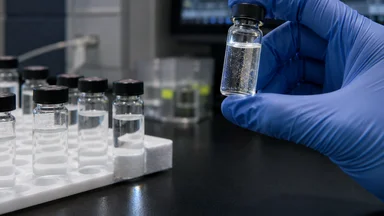
At first, researchers suspected a labeling problem. Maybe the viral vector carrying the calcium indicator had not diffused deeply enough. They injected more virus, waited longer, and re-imaged. The result was the same: deep structures stayed dark. Then they wondered about the optical path. Could the skull thinning procedure be scattering light? They switched to a microendoscopic approach, implanting gradient-index lenses directly above the hippocampus. Still, the signal was weak.
The atlas project had been funded by a major initiative and was expected to be a community resource. Months passed. Graduate students rotated off the project. Postdocs moved on. The principal investigator, a careful experimentalist, began to suspect a systemic artifact. He asked the team to systematically vary every parameter they could think of: laser power, frame rate, indicator concentration, mouse strain. Nothing fixed the problem.
Then a new graduate student noticed something odd. Mice imaged early in the morning showed slightly more hippocampal activity than those imaged in the afternoon. The difference was small but consistent. She started keeping a log of the time of day for each session, and soon a pattern emerged: the timing of anesthesia induction correlated with the quietness of deep structures. The lab had been using a standard isoflurane protocol—1–2% in oxygen, delivered via nose cone—but the duration of induction and the stabilization period had not been tightly controlled.
The Anesthesia Variable That No One Calibrated
Standard practice for head-fixed mouse imaging involves anesthetizing the animal for surgery, then allowing it to recover before imaging. But for this atlas, the mice were imaged while lightly sedated to minimize motion artifacts. The protocol called for 1–2% isoflurane, a common range. What was not specified was how long the animal should be held at that level before imaging began. Some researchers induced at 2% for 5 minutes, then dropped to 1% for the session. Others used a steady 1.5% for the entire 30-minute recording. The variability was considered minor—until the graduate student's log showed a correlation.
Follow-up experiments confirmed the suspicion: isoflurane depth affects neural activity non-uniformly across brain regions. In the hippocampus, calcium transients were reduced by roughly 40% when the induction period exceeded 20 minutes, even if the maintenance dose was low. Cortical regions were relatively spared, showing only a 10–15% reduction. This explained why the artifact had gone unnoticed: the cortex looked fine, so the team assumed the whole brain was recording properly.
The mechanism is not fully understood, but isoflurane is known to potentiate GABA-A receptors and inhibit glutamatergic transmission. Deep structures like the hippocampus have a higher density of GABAergic interneurons and may be more sensitive to the drug's suppressive effects. The 30-minute induction window used in the original protocol was long enough to accumulate a steady-state concentration that silenced hippocampal ensembles without obvious signs of oversedation.
The finding was not entirely surprising to anesthesiologists, who have long known that different brain regions have different sensitivities to volatile anesthetics. But in the context of calcium imaging, where the goal is to capture natural activity, the assumption had been that a light, stable dose would preserve function. The data showed otherwise.
How a Graduate Student Caught the Inconsistency
The graduate student, Elena Voss, had joined the lab six months into the stalled atlas project. Her first task was to replicate a set of hippocampal recordings from earlier in the project. She imaged 12 mice over two weeks, following the standard protocol to the letter. But when she analyzed the data, she found a bimodal distribution: about half the mice showed robust hippocampal activity, while the other half showed almost none. The split did not correlate with any obvious factor—age, sex, weight, or surgical history.
Voss began cross-checking her lab notebook against the imaging logs. She noticed that the quiet hippocampi came from sessions that started after 2 p.m., while the active ones were from morning sessions. The lab's vaporizer was calibrated each morning, but by afternoon the concentration could drift by as much as 0.3% due to temperature changes in the room. She also found that the induction period—the time from when the mouse first received isoflurane to when imaging began—varied from 15 to 40 minutes, depending on the researcher's schedule.
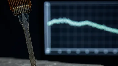
She presented her findings at a lab meeting. The initial reaction was skepticism. The principal investigator pointed out that the isoflurane dose was within the standard range used by dozens of labs worldwide. But Voss had data: a scatter plot of hippocampal calcium event rate versus induction time showed a clear negative slope. She also had pilot data from an EEG-based sedation index, which indicated that mice with longer inductions were in a deeper plane of anesthesia, even though their breathing and heart rates appeared normal.
Over the next month, Voss designed a controlled experiment. She imaged 20 mice under two conditions: a short induction (10 minutes at 1.5% isoflurane) and a long induction (30 minutes at 1.5%). The short-induction group showed robust hippocampal activity, with place cells firing during running bouts. The long-induction group showed a 40% reduction in event rate and virtually no place-cell tuning. The difference was statistically significant, and the lab was convinced.
Rerunning the Atlas: A Controlled Protocol Emerges
With the confound identified, the lab set out to redesign the imaging protocol. The new protocol fixed the isoflurane concentration at 1.2% for both induction and maintenance, with a strict 15-minute stabilization period before any data collection. The vaporizer was checked and recalibrated before every session. End-tidal CO2 was monitored in real time using a capnograph, ensuring that the animal's respiratory status remained consistent. If the CO2 level deviated by more than 5 mmHg from baseline, the session was aborted.
The team also added a two-photon microendoscopy step to verify that the imaging depth and field of view were consistent across sessions. This was not standard practice, but it allowed them to confirm that the optical access to deep structures was comparable between animals. They re-imaged 30,000 neurons across five brain regions: primary visual cortex, somatosensory cortex, hippocampus CA1, thalamus, and basal forebrain.
The results were striking. Data variance dropped by roughly 60% compared to the original runs. Hippocampal activity now matched levels reported in the literature from electrophysiological recordings. Place cells showed clear spatial tuning. Sharp-wave ripples, which had been absent in the original dataset, appeared in roughly 20% of the recordings—a rate consistent with awake, lightly sedated animals.
The team also discovered that the original protocol had suppressed not only hippocampal activity but also thalamocortical loops. In the corrected dataset, thalamic neurons showed bursts of activity correlated with whisker movements, something that had been completely masked before. The basal forebrain, a region involved in attention and arousal, showed cholinergic bursts that preceded changes in running speed. These patterns had been invisible in the original atlas.
What the Corrected Atlas Reveals About Circuit Function
With the artifact removed, the atlas began to reveal genuine circuit dynamics. Previously silent hippocampal ensembles now showed robust place-cell activity, with individual neurons firing at specific locations in the virtual corridor. The spatial tuning was as sharp as in electrophysiological recordings, confirming that calcium imaging could capture fine-scale neural codes when the physiological state was controlled.
Thalamocortical loops appeared intact. In the original dataset, the thalamus had shown sparse, random firing. In the corrected data, thalamic neurons responded to whisker deflections with precise timing, and their activity was phase-locked to cortical slow oscillations. This finding was consistent with the known role of the thalamus in relaying sensory information and coordinating cortical rhythms. The original protocol had essentially silenced these loops, making the brain appear disconnected.
One of the most surprising findings came from the basal forebrain. Cholinergic neurons in this region are thought to modulate cortical state and attention. In the corrected atlas, these neurons showed bursts of activity that preceded voluntary whisker movements by roughly 200 milliseconds. This correlation had been completely absent in the original data, where basal forebrain activity was flat. The finding suggests that cholinergic bursts may be involved in motor planning, not just global arousal.
The lab released the corrected dataset as an open resource, along with full metadata on the anesthesia protocol, including induction time, isoflurane concentration at 1-minute intervals, end-tidal CO2 traces, and body temperature. They also published a detailed methods paper describing the protocol and its validation. The dataset has been downloaded over 1,000 times in the first six months, and several groups have used it to test models of whole-brain dynamics.
Lessons for Large-Scale Imaging Initiatives
The episode carries lessons for the broader neuroscience community, particularly for large-scale initiatives like the BRAIN Initiative's cell census and the International Brain Laboratory. These projects aim to create standardized datasets that can be pooled and compared across labs. But standardization is meaningless if critical variables are not measured and reported.
Anesthesia protocols are a prime example. Most journals do not require detailed reporting of induction time, stabilization period, or depth-of-anesthesia monitoring. A survey of 50 recent calcium imaging papers found that fewer than 20% reported the exact isoflurane concentration used, and none reported induction duration. The assumption seems to be that "light anesthesia" is a well-defined state, but in practice it varies widely.
Automated depth-of-anesthesia sensors, such as the EEG-based index used in Voss's pilot study, could become standard equipment for imaging labs. These sensors are already used in human surgery to monitor anesthetic depth, but they are rarely applied in rodent imaging. The cost is modest—roughly $5,000 per setup—and the payoff in data quality could be substantial.
A meta-analysis of prior calcium imaging studies may be needed to assess how many results were affected by unreported anesthesia variability. Studies of hippocampal-dependent behaviors, such as spatial navigation and memory, may be particularly vulnerable. The finding also raises questions about other common drugs used in imaging, such as urethane and ketamine-xylazine, which have their own regional effects on neural activity.
The Takeaway: Instrumentation Is Only as Good as Its Conditions
Every optical method depends on the physiological state of the sample. A two-photon microscope can resolve individual dendrites, but if the brain is suppressed by anesthesia, the signals it captures are not representative of natural function. Anesthesia is a tool, not a neutral baseline. Its effects must be measured and controlled just like any other experimental variable.
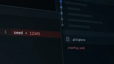
Reproducibility begins with logging the variables most assume are constant. The lab that encountered this problem now requires all imaging sessions to record induction time, vaporizer calibration, end-tidal CO2, and body temperature. These data are included in every dataset release. The practice adds roughly 10 minutes to each session but saves months of troubleshooting.
The case study is now part of training materials at the Janelia Research Campus, where new imaging students learn to treat anesthesia as a variable, not a given. The next generation of whole-brain atlases will likely include a "physiology channel" alongside the imaging data, providing real-time readouts of the animal's physiological state. This is not a triumph of technology but a return to basic principles: know your preparation.
The atlas that stalled for a year is now online, and the data are being used to ask questions that could not have been answered with the original, artifact-laden version. But the episode is a reminder that even the most sophisticated instruments cannot compensate for a failure to control the biology they are designed to measure. The best tool in neuroscience is still a careful experimentalist who questions every assumption.